Radiologists use several imaging techniques for diagnosing breast cancer. One common method is magnetic resonance imaging – or MRI. A breast MRI combines radio waves and magnetic fields to create detailed images of the breast on a computer. Discover more about this testing procedure and what it reveals from the breast and women’s health medical team at Richmond University Medical Center in Staten Island, NY.
How Does a Breast MRI Work?
The purpose of this type of MRI is to identify cancer or other irregularities in the breast. Typically, a patient undergoes an MRI after receiving a positive result during a breast cancer biopsy.
Patients with a high risk of breast cancer may go through an MRI in conjunction with a mammogram to screen for cancer. Mammography is not always successful at spotting breast cancer in younger women or those with breast implants. Additionally, studies show that MRIs can find small breast lesions that mammograms sometimes overlook.
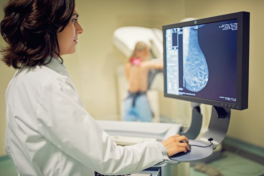
To begin the procedure, the technician may inject a contrast dye called gadolinium into the patient’s arm to make the results easier to read. The patient will lie face down with their breasts resting in the openings on the table. Once the patient is properly positioned, the table slides into the MRI machine and the test begins.
While administering the test, the technician will sit in another room with a window, monitoring breast placement and possible movement. In most cases, the technician will have a microphone that enables them to deliver instructions to the patient. That connection also allows the patient to express any concerns throughout the procedure. The entire examination generally takes around 30 minutes to an hour.
When Is a Breast MRI Administered?
Breast MRIs help physicians understand the extent of cancer in a patient. It is frequently used to detect the disease in patients believed to be at high risk for breast cancer, but a provider may also order the test for the following other reasons:
- To further assess abnormalities found during an ultrasound or mammography
- To decide whether lumpectomy or mastectomy is the most effective treatment option
- To detect breast cancer early in women with dense breast tissue
- To look for cancer in the other breast not recently diagnosed
- To assess a newly inverted nipple change
- To evaluate the leakage of a silicone gel breast implant
- To check for cancer recurrence following a lumpectomy
- To see if cancer expanded into the chest wall, which may alter the treatment
- To examine the size and location of breast cancer lesions
- The patient went through radiation treatment in the chest between the ages of 10 and 30
- The patient has a history of precancerous breast changes (lobular carcinoma in situ or atypical hyperplasia)
- The patient has implants or scar tissues that may interfere with an accurate mammogram
- The patient or a close relative (mother, daughter, or sister) has a BRCA1 or BRCA2 gene mutation
- The patient or a close relative has Bannayan-Riley-Ruvalcaba syndrome, Cowden syndrome, Li-Fraumeni syndrome, or a similar genetic disorder
About Breast MRI Test Results
After the test, a radiologist analyzes the images. These health professionals use the same system to discuss breast MRI results as they do mammograms and ultrasounds. This system – called the Breast Imaging Reporting and Data System (BI-RADs) – categorizes the outcomes ranging from zero to six. This approach simplifies communication about results for all three testing methods.
Once the radiologist shares the results with the patient’s primary care provider, the physician will explain what they mean and how they may affect breast cancer treatment going forward.
Seek Breast Imaging Services at Richmond University Medical Center
Breast MRIs are vital for patients, especially if they received unreliable results from a mammogram or ultrasound. Those in need of breast imaging in the Staten Island, NY, area should turn to Richmond University Medical Center for compassionate and efficient care. At the Breast and Women’s Center, patients can obtain several examinations, from routine mammograms to breast biopsies.
A non-profit healthcare provider, Richmond University Medical Center fosters a positive environment to ensure patients, families, physicians, and staff members experience the highest satisfaction possible. Contact us today to learn more about breast MRIs and other care services.